Reproduction of David Maurice web site at Columbia [ DavidMaurice.org home page ]


 The Penetration of Drugs from the Tears to the Retina

  D Maurice, S Velilla, anb L-S Chuang

Columbia University Department of Ophthalmology

presented at  ARVO, 1999


SUMMARY:
Usually, studies of the penetration of a drug from the tears to the retina and vitreous are carried out in the rabbit, somewhat as follows:

Standard Technique

  • Topical instillation of drug
  • Animal killed after specified period
  • Conjunctival sac flushed
  • Aqueous collected
  • Conjunctiva collected
  • Globe enucleated
  • Vitreous, retina collected
ERRORS IN STANDARD TECHNIQUE
1. Contamination
Tracers of drug from lid margins etc. can spread onto surface of globe during enucleation and then into vitreous.

2. Penetration After Death
Choroidal blood flow maintains zero concentration at the RPE and should be a major obstacle to drug penetration.
   When the flow ceases at death there are about 5 minutes before the retina is collected during which a drug concentration should build up at the RPE.

AVOIDANCE OF ERRORS
In the present experiments we attempted to avoid these errors by:

1. Small subconjunctival injection
This provides a circumscribed zone of high penetration with a border free of drug.

2. Freezing eye after death
Suppresses postmortem diffusion and spreading of external contamination.

EXPERIMENTAL PROCEDURES
  •  Rabbit anesthetized
  •  Subconjunctival injection of fluorescent tracer
  •  Wait 5 - 75 minutes
  •  Animal killed with i.v. pentobarbital
  •  Eyes frozen in liquid nitrogen either immediately or after 5 min delay
  •  Decapitation
  •  Core of frozen globes machined out
  •  Core lyophilized
  •  Dried specimens separated into component tissues
  •  Tracer extracted from tissues and measured
TRACERS
   Dyes were used as tracers because their distribution after injection is visible. Fluorescent dyes can be determined in very low concentrations.
   Rhodamine B (RB) and Sulforhodamine B (RSB) were chosen for representing extremes of lipophilicity.
 INJECTION
 A measured volume, ~ 0.2 µl, of 10% SRB or 1% RB injected RE through glass electrode near equator nasal to superior rectus.  The solution was made viscous with hyaluronic acid to prevent regurgitation.
FREEZING
The animal is elevated on a platform.
After death, the right eye is proptosed (A) and dipped into liquid nitrogen (B) for about 1 min. The left eye is then proptosed (C) and both eyes plunged into liquid nitrogen (D) until boiling ceases.
PREPARATION OF CORE
To firmly anchor the frozen head a 1/4” hole is drilled through it and a bolt is passed through it.

The bolt is fixed in a yoke and attached to the tool post of a hobbyists’ lathe.

A 16 mm diameter channel is bored into the globe with a chilled tool.

The core is separated with a saw.
 

SEPARATION OF TISSUES
The core is lyophilized for 24 hr.

The vitreous can be lifted out cleanly in one piece.

The retina comes off in flakes on pressing with a tool. The RPE sometimes comes with it, sometimes not.

The choroid is scraped off the sclera.

A colored spot is seen centrally on the inside of the sclera of injected eyes. It becomes spread out in the longer term experiments.

The sclera can be split form conjunctival tissue.

The fluorescent label is extracted from the tissue samples and measured in a fluorometer that can estimate 1 picogram.
 

RESULTS
  • The results for RB and SRB are similar except that the penetration of the latter was an order of magnitude smaller.
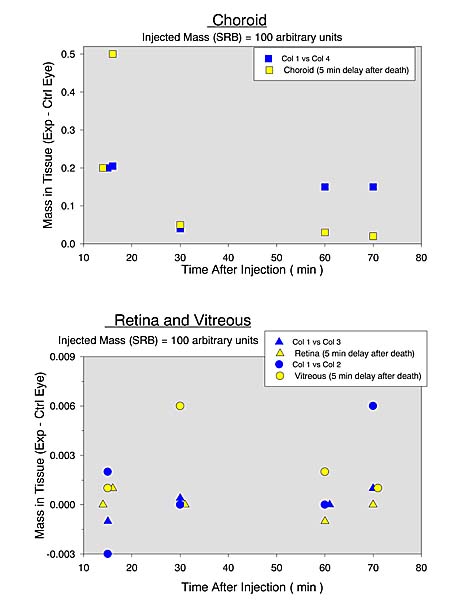
  • The amount of label in the vitreous and retina of the control (left) eye, is generally of the same order of magnitude as that in the right eye. The differences between the two eyes are plotted.
  • The amount of label in the choroid of the right eye is considerably raised.
  • There is no marked increase in the penetration into any tissue as a result of a 5 minute postmortem delay before freezing.
  • The total mass of tracer that can be eluted from the sclera and conjunctiva falls off very slowly with time.
Rhodamine B
Conjunctiva + Sclera
Rhodamine B
Choroid


Rhodamine B

Retina and Vitreous
Sulforhodamine B
Conjunctiva + Sclera
Sulforhodamine B
Choroid


Sulforhodamine B

Retina and Vitreous
DISCUSSION
  • The absence of an increase in the fluorescent tracer in any of the tissues as a result of the 5 min postmortem period is surprising.

  • In the retina and vitreous this could be explained if the RPE was a very large barrier.

    In the choroid it perhaps could be accounted for in part if the tracer accumulated in the stagnant blood in the choroidal vessels and this was squeezed out of the eye during freezing. 

    Further investigation is needed.

  • From their appearance on the inner surface of the sclera it seems that the tracers can diffuse readily both across and in the plane of the tissue. It is strange therefore that they are not lost more rapidly to the conjunctival and choroidal circulation.
CONCLUSIONS
  • The penetration of a lipophilic tracer RB into the vitreous and retina is of the order of 0.01% and of a hydrophilic tracer SRB of the order of 0.001% of the amount in a subconjunctival dose.

  • It is to be expected that therapeutic drugs with similar physicochemical properties would behave similarly.

  • In conjunction with preliminary studies of the penetration of tracers from the tears to the sclera, not reported here, the present findings offer no support for the hope that a therapeutic concentration can be reached in the posterior segment by the topical application of a drug.
REFERENCES:

Supported by NEI and Research to Prevent Blindness


[ David Maurice Home Page ]

Reproduction of David Maurice web site at Columbia [ DavidMaurice.org home page ]