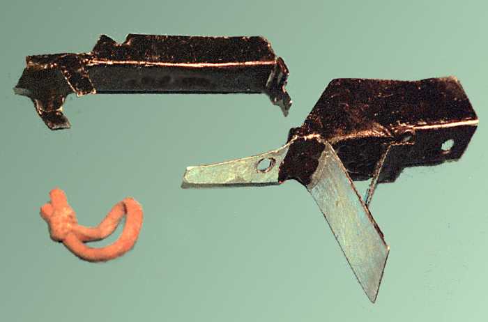
| CONSTRUCTION |
|

  |
| Reproduction of David Maurice web site at Columbia | [ DavidMaurice.org home page ] |
| INTRODUCTION |
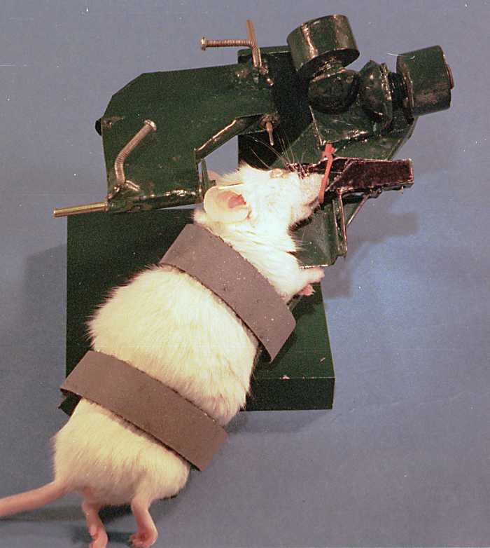
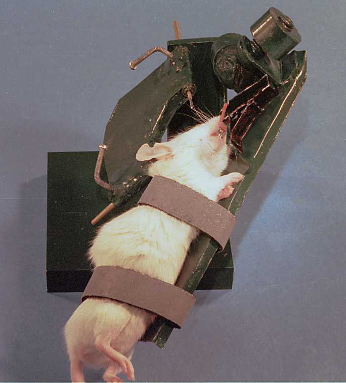
This is facilitated by being able to rotate the animal around the center point of the eyeball in a system of gimbals, so that the cornea remains in focus as the area under observation is shifted. |
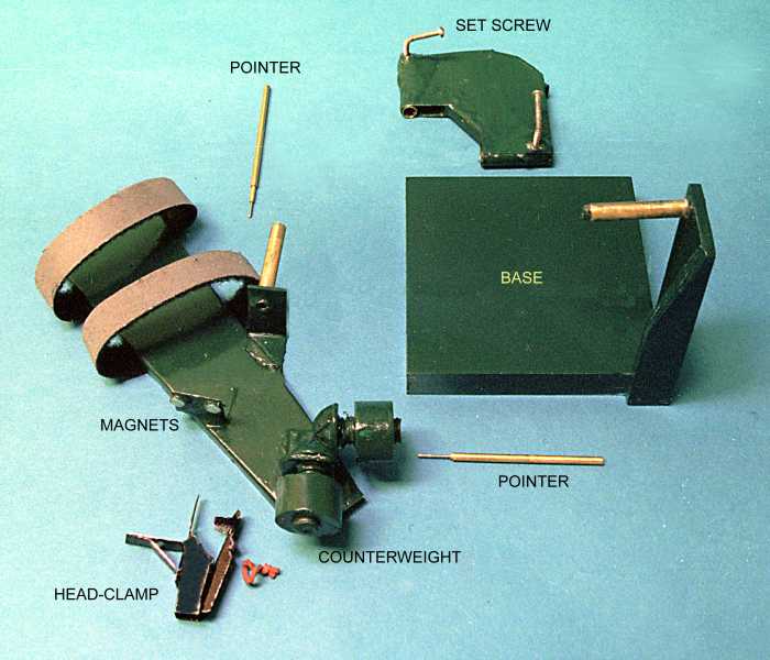
| CONSTRUCTION |
|
|
|
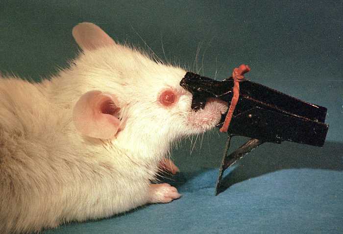
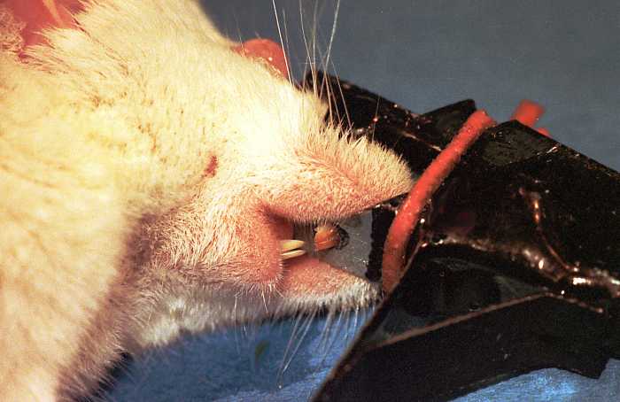
| OPERATION |
|
    |
|
|
|
| Reproduction of David Maurice web site at Columbia | [ DavidMaurice.org home page ] |